- Contact Person : Mr. Zhang Hyde
- Company Name : Wuhan Jingcheng Weiye Medical Equipment Co., Ltd.
- Tel : 86-027-88034149
- Address : Hubei,Wuhan,xiongchu Road
- Country/Region : China
- Zip : 430000
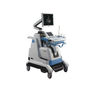
Newest Color Doppler Ultrasonic Diagnostic System Q6 with CE certificate


Related Product Searches:Newest Color Doppler Ultrasonic Diagnostic System Q6 with CE certificate,High Quality,Color Doppler Ultrasonic Diagnostic System, Intelligent diagnostic system,Q6
System OverviewApplicationAbdomenCardiacObstetricsGynecologyUrologySmall PartsVascularPediatrics
Electrical PowerVoltage: 100V~240VCurrent: 7.0A/4.0AFrequency: 50/60Hz
Physical SpecificationsEquipment:910mm×660mm×1470mm; 68kgPackage:1035mm×725mm×1535mm; 128kg
ConditionsOperatingTemperature: 10°C~40°CHumidity: 30%~75%Pressure: 50kPa~106kPaShipping/StorageTemperature: -15°C~50°CHumidity: 10%~90%Pressure: 50kPa~106kPa
Connectivity/Media/PeripheralsTransducer Ports: 3 (Activated)USB Ports: 5Hard Disc: ≥ 160GBFootswitch: BNC/USBEthernet Port: 1 (10Mb/1000Mb)External Display: DVI, VGAExternal VCRDigital Video MonitorCompound Video OutputBarcode ReaderUSB ModemUSB PrinterPeripherals/Storage TrayDigital B/WThermal PrinterRemote Diagnosis ModuleDVD/CD Recorder
Image StorageStorage Format:PNG, AVI, MPEG, RAW(cine)Export Video Format:AVI, MPEG, DICOMExport Image Format:PNG, JPEG, BMP, GIF, DICOMCine Frame Capacity:> 8000 framesStored Image Capacity:> 50000 imagesCD-R StorageDVD Storage
TechnologyTP-view TechPanoramic Imaging TechSpeckle Reduction TechDual & Triplex Synchronous DisplayElastography TechDirectional Power DopplerReal-time 3D Volume Imaging TechExpand-Pulse-Inversion Imaging TechColor Deflection TechFree-hand 3D Imaging TechSpatial-Compound Imaging TechPulse-Inversion Harmonic Imaging TechImaging Parameters PresetCustom Key/Optimization Key
ECG (Optional)ECG ModuleECG LeadECG InvertECG BaselineECG Gain
Embedded ComputerCPU:Intel E4300 (Minimum)Hard Drive (Patient) Data:≥ 500 GB
Hardware SpecificationLCD MonitorSize (Diagonal): 15”Contrast Ratio: 1500:1Resolution: 1024×768 pixelsBrightness: 250 cd/m2Color Depth: 24bitRotate Angle: ±90°Grey Levels: 256Touch ScreenSize (Diagonal): 8.4”Weight: 330gContrast Ratio: 600:1Resolution: 1024×768 pixelsBrightness: 450 cd/m2Grey Levels: 64Input Signal: LVDSCompatible with bare hands/thin medicalgloves (e.g., latex, vinyl, nitrile)
Software LanguagesEnglishGermanSpanishItalianCzechFrenchRussianSimplified ChineseBrazilian Portuguese
Imaging PerformanceStartup Time (Max):Avg. < 90 secondsPreset Switching Time:Avg. < 1 secondStorage Time (Image to Disk):Avg. < 0.5 second
TransducersConvex Array ProbeFrequency:Central 3.3 MHzAverage Min. 3.15 MHzMax. 3.85 MHzPitch: 0.468 mmRadius: 60 mmNumber of Elements: 128
Geometric Focus Approx: 90 mmMicro-Convex Array Probe:Frequency:Central 4 MHzAverage Min. 3.6 MHzMax. 4.4 MHzPitch: 0.28 mmRadius: 20 mmNumber of Elements: 128Geometric Focus Approx: 90 mmLinear Array Probe:Frequency:Central 10 MHzAverage Min. 7.2 MHzMax. 8.8 MHzPitch: 0.390 mmRadius: N/ANumber of Elements: 128Geometric Focus Approx: 30 mmIntra-Cavity Probe:Frequency:Central 6.5 MHzAverage Min. 5.85 MHzMax. 7.15 MHzPitch: 0.205 mmRadius: 10 mmNumber of Elements: 128Geometric Focus Approx: 35 mmPhased Array Probe:Frequency:Central 2.5 MHzAverage Min. 2.25 MHzMax. 2.75 MHzPitch: 0.300 mmRadius: N/A
Number of Elements: 64Geometric Focus Approx: 30 mm3D/4D Volume Probe
Imaging ModesOverviewB, Dual B, Quad BM, Color M, Anatomical MColor, Dual Color, Simultaneous 2D/ColorCompound, Dual CompoundPW, CW, TriplexCFM, PD, Directional PD, CDFreehand 3DStandard 3D/4D, Advanced 3D/4DPanoramic, ElastographyElastography ComparativeTissue Doppler Imaging (TDI)Expand Pulse Imaging (EPI)
Combined ModesDual B, Quad B, B/M, B B/C, Dual B/C
B/PW, B/CW, B B/Power, Dual B/PowerDual Compound, B/Elasto, B B/ElastoB B/TDI, Dual B/TDIB/C/M, B/C/PW, B/C/CWB/Power/M, B/Power/PW, B/Power/CWB/TDI/M, B/TDI/PW, B/TDI/CW
Standard 3D/4D ModeMaps: 17Line Density: 64~192Rendering Quality: Low, Med, HighFrames per Volume: 3~125Field of View (Max): 5°~75°Viewing Options: A/B/C/VR, A/VR,C/VR, VRendering Methods: Surface, Surface-Smooth, Maximum, X-RayOne Sweep 3D, 3D ROI, 3D Niche Mode,Measurements, Sculpt, Annotations,Contrast, Threshold
Advanced 3D/4D ModeMaps: 17Line Density: 64~192Rendering Quality: Low, Med, HighFrames per Volume: 3~125Field of View (Max): 5°~75°Max Volume Rate: 3Viewing Options: A/B/C/VR, A/VR,C/VR, VRendering Methods: Surface, Surface-Smooth, Maximum, X-RayLive 3D/4D imaging, One Sweep 3D,Rendering 3D ROI, Multi-Slice,3D Niche Mode, Measurements,Sculpt, Annotations, Contrast, Threshold,Save Cine-loops, Save Volume
Analysis PackagesOBCardiacAbdominalGYNUrologyVascularSmall Parts
Standard PackageMain Frame (with a 15”LCD Monitor)Customers can select any 2 probesaccording to their needs from Convex,Intra-cavity, Linear and Phased ArrayProbes.Power cord/grounding cableFuse (2)User’s ManualCoupling Gel (0.25L)
Optional Accessories17”LCD Monitor4D Imaging ModuleElastography ModuleConvex Array ProbeMicro Convex Array ProbeLinear Array ProbePhased Array Probe4D Volume ProbeIntra-cavity ProbeDICOM 3.0Pedal SwitchLaser, Ink-jet PrinterPuncture TrestleBarcode ReaderECGUPS
CertificatesQuality StandardsISO9001ISO13485CMDCE Certificate
Newest Color Doppler Ultrasonic Diagnostic System Q6 with CE certificate